
犬焦虫症与检验技术
焦虫的发现
焦虫症 (babesiosis),或称为巴贝氏虫症、壁虱热(tick fever)、德州热(Texas fever),是由许多不同品种的焦虫属 (Babesia sp.) 原虫寄生于红血球内所引起之疾病,会感染各种脊椎动物,包括家畜、野生动物及人等等。这些原虫属于梨形虫目(Piroplasmida)、巴贝氏科(Babesiidae)、巴贝氏虫属(Babesia),全世界超过100 种。是19世纪末时,由罗马尼亚的外科医生Victor Babes在观察牛跟羊血红素尿 (hemoglobinuria) 的红血球细胞时,所发现的一种新型微生物,后来分别被命名为牛焦虫 (Babesia bovis) 与羊焦虫 (Babesia ovis) [1]。不久后,1895年就在犬的身上,也发现焦虫感染的纪录 [2]。直至今日,世界各地都有发生过焦虫感染的纪录[4]。
焦虫分类
传统上,会利用显微镜观察血液抹片下红血球中梨形裂殖子 (piroplasm merozoites)
的型态来分类大焦虫和小焦虫。而现在利用新兴分子生物学技术,让科学家可以鉴定出更多种造成犬只感染焦虫症的病原体。原先专指B. canis的大焦虫,也被发现有Babesia rossi 、 Babesia vogeli 等新的品种。形态上,B. rossi、B. vogeli 两者完全相同使得它们被认为是B. canis 的亚型。但在临床表现、地理分布、媒介特异性的显著差异[3],有人认为其实这三者分属于不同的品种。另外,在北美洲发现的Babesia bigemina也被认为属于大焦虫类 [4-8 ]。小焦虫类目前只有三种被记录,分别是B. gibsoni、B. conradae [9, 10] 和B. vulpes [11]。其中Babesia vulpes在先前报导被描述为"Spanish dog isolate"、 “Babesia microti-like"、 "Babesia (Theileria) annae" 或 “Babesia cf. microti”。因此,随着分子生物技术的进步,我们将有机会发现更多品种的焦虫。
犬焦虫症的临床症状
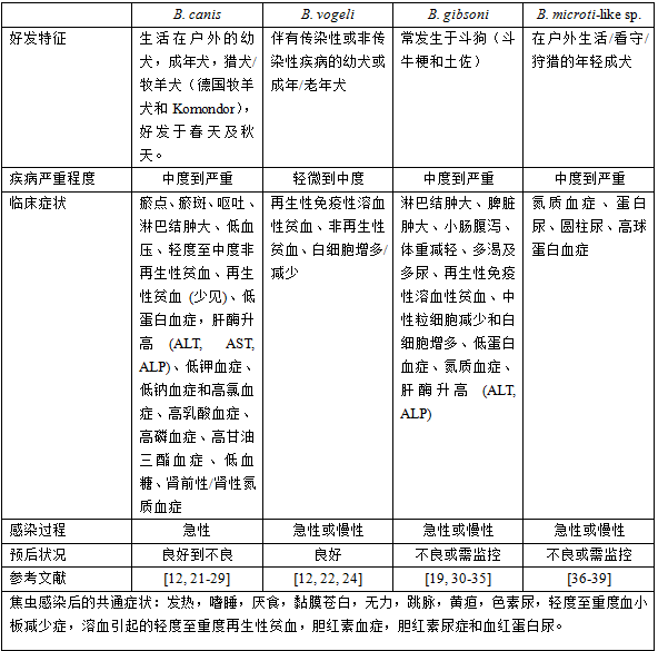
感染焦虫症的宿主依照其免疫力,及虫体的血清型而有不同表现。一般共同的症状有倦怠、虚弱、厌食、黏膜苍白、发烧、淋巴结和脾脏肿大、贫血、血小板减少、黄疸和色素尿 (pigmenturia)。尽管经常检测到不同程度的血小板减少症状,但是瘀点或瘀斑的存在则不太常观察到。感染时,血小板减少与贫血症状从轻微到严重都可能发生。其他可以检测到的异常还包括低蛋白血症 (hypoalbuminemia) 和高胆红素血症 (hyperbilirubinemia) [4, 8]。感染焦虫后的贫血是由于焦虫引起的损伤和红血球破裂引起的血管内和血管外溶血以及细胞渗透脆性增加。一般而言,焦虫感染的贫血为再生性贫血,但由B. canis造成的严重贫血也可能会变成非再生性贫血 [12]。许多狗可能出现其他临床症状,这些症状与感染后造成的溶血没有直接关系,但表示其他器官也受到影响,尤其是被B. rossi感染后症状最为明显,包括体重减轻、急性或慢性肾病、肾小球肾炎、凝血功能障碍 (瀰漫性血管内凝血) 、血液浓缩、休克、代谢或呼吸性硷 (酸)中毒、胃肠道疾病 (呕吐或腹泻) 、胰腺炎、腹水、眼部病变 (葡萄膜炎或失明) 、肌肉疼痛、横纹肌溶解症和呼吸系统疾病 (水肿或急性呼吸窘迫) [13]。感染焦虫后可能发生的症状众多,不同种的焦虫症状又稍有差异。根据各国实验室收集的众多资料,将感染的特征、过程与预后整理如表一。
犬焦虫症的检验方式
一、显微镜观察
血液抹片检查是犬焦虫病的诊断工具之一。利用显微镜下观察是最容易和方便的诊断测试。然而,这种方法的灵敏度低于分子诊断技术,同时也取决于每只犬感染状况的不同。利用血液抹片检查最好是使用新鲜的血液。另外,如果要检验大焦虫,从耳朵或是指甲采集毛细管血管的血可以得到较多的大焦虫,提高检验的灵敏度 [8,14]。根据文献,以显微镜观察血液抹片的检测极限约为0.5% [14]。图一是大焦虫类与小焦虫类的血液抹片,在显微镜下观察的状况。
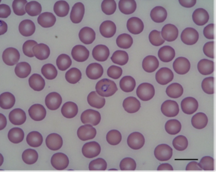
图一 小焦虫 (B. gibsoni)的血液抹片
利用显微镜观察血液抹片来检查是否感染大焦虫是兽医师很常见的做法,但是要分辨出像B. vogeli这类的亚型就比较困难,此时就必须利用像PCR等其他方法来协助区别。至于用显微镜观察小焦虫 (B. gibsoni, B. microti-like sp.) 感染,由于形态上的缘故,鉴别上就更加困难,需要较有经验的医师才能得到较好的检验灵敏度。显微镜观察血液抹片应该做为焦虫感染筛检的第一步,当显微镜观察未发现焦虫感染时,可以利用更具敏感性的方法如PCR来确认为感染阴性。国外期刊建议检验焦虫的流程如图三所示。
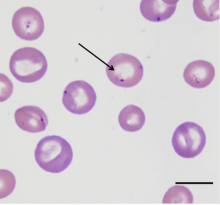
图二 大焦虫 (B. canis)的血液抹片
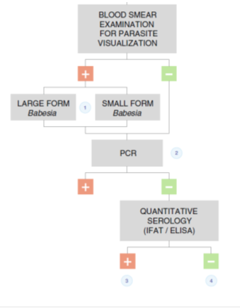
图三 焦虫的建议检验流程
二、血清学检验
酶联免疫吸附法 (ELISA) 或间接免疫荧光法 (IFAT) 是常用的定量式血清清学检验技术。IFAT或ELISA的优点是可以定量血清中抗体的高低,数值化可以协助判断犬只患病/预后的进程 [40]。目前市面上已有根据ELISA原理所开发的快速检验试剂,可以分辨出血液样本中是否含有感染后所产生的抗体。不过这些检测试剂多半只能分辨有或无,无法定量抗体的高低。因此一般而言,进行血清学检验时,大多还是送至专门的实验室检验。此外,实验室进行的检验在灵敏度与特异性上也较快速检验试剂更好,快速检验试剂还是较适宜做为初步筛检使用。
临床上,以血清学方式检验焦虫感染要注意感染的期程。尤其如B.canis常见为急性感染,有可能发生犬只已经感染,但血清为阴性的空窗期 [4, 41]。因此血清转化 (seroconversion) 可用来做为焦虫血清学检验的一个指标。感染后3-4周产生的阳性抗体力价为中到高,可确认焦虫的感染时期 [37]。正常情况下,当疑似患病的犬只出现临床症状或实验室检验异常时,可进行初步血清学检验,待4-8周后进行再次血清学检验复验。可惜目前关于血清转化的临床数据报告仍不够多,这部分需要更进一步的累积资料,才能得到更具临床意义的血清转化时间点。
对于焦虫检测,使用血清学检验可能会遇到不同焦虫感染抗体交叉干扰的状况,尤其是型态/亲缘关系相近 (phylogenetically related) 的品种间,如大焦虫B. canis、B. vogeli、 B. rossi之间与小焦虫B. gibsoni、B. microti-like sp之间都可能会互相干扰 [42]。虽然复数感染的发生机率并不高,但是血清学检验还是用来诊断目前或曾经感染焦虫的状况为佳。区分焦虫的品种建议使用分子生物学技术来判断。
三、分子生物技术
一般而言,PCR在诊断焦虫感染时有几个优点。首先,PCR检测比直接血液抹片检查更加灵敏,再者,能够直接侦测到病原特定的DNA片段,可以视为一个病原仍相当活耀的证据,代表犬只目前持续感染中。此外,与光学显微镜或血清学直接检测不同,PCR可以更准确地识别致病的品种 [4]。许多新的分子技术都用来鉴定和区分各种焦虫,包括半巢式 (semi-nested) PCR [15],反向线点PCR (PCR-based reverse line blot) [16, 17]和PCR限制性片段长度多态性分析 (PCR-restriction fragment length
polymorphism analysis) [18]。常用来区分不同种焦虫的DNA片段为rRNA (nuclear ribosomal RNA ) [5, 6] 和两个内部转录间隔区(ITS1和ITS2)[5].
利用这些分子技术检验,能够有效地区分犬只是被何种焦虫感染,进而提供更准确的治疗,也能有效追踪预后状况。许多犬只长期感染焦虫,一直处于慢性感染状态下,会使得这些犬只处于易于复发或维持长期异常的状况。但利用血液抹片观察又无法有效判断虫体是否存在。此时,PCR就可用来确定感染是否仍然存在或已经被有效清除 [4,8]。追踪预后状况应在中断治疗前和治疗结束后约2个月进行,特别是被小焦虫感染的犬只,更应该追踪。尽管 PCR 检验灵敏度极高,侦测范围仍然有其极限,建议可以进行两次连续PCR检测,间隔至少15天 [19, 20]。此外,在追踪时,除了采取外围液血检测之外,也使用多种组织抽吸物进行PCR,例如脾脏组织,才能严格确认寄生虫的清除效果 [8]。
结论
近年对于焦虫的研究越来越多,使得我们对于犬焦虫症有更多的了解。由于焦虫症是有多种类焦虫感染的疾病,因此准确的检测、正确的辨识焦虫种类,才能有效的选择最适合的治疗方法以及做好预后照顾。
表一 感染不同种类焦虫的犬只的主要临床表现和预后 (资料所在地:欧洲)
参考资料:
1.Uilenberg G. Babesia-a historical overview. Vet Parasitol. 2006;138:3–10.
2.Roncalli AR. The history of Italian parasitology. Vet Parasitol. 2001;98:3–30.
3.Carret C, Delbecq S, Labesse G, Carcy B, Precigout E, et al. Characterization
and molecular cloning of an adenosine kinase from Babesia canis rossi. Eur J
Biochem. 1999;265:1015–21.
4.Solano-Gallego L, Baneth G. Babesiosis in dogs and cats – expanding
parasitological and clinical spectra. Vet Parasitol. 2011;181:48–60.
5.Zahler M, Schein E, Rinder H, Gothe R. Characteristic genotypes discriminate
between Babesia canis isolates of differing vector specificity and pathogenicity
to dogs. Parasitol Res. 1998;84:544–8.
6.Carret C, Walas F, Carcy B, Grande N, Precigout E, et al. Babesia canis canis,
Babesia canis vogeli, Babesia canis rossi: differentiation of the three
subspecies by a restriction fragment length polymorphism analysis on amplified
small subunit ribosomal RNA genes. J Eukaryot Microbiol. 1999;46:298–303.
7.Caccio SM, Antunovic B, Moretti A, Mangili V, Marinculic A, et al. Molecular
characterisation of Babesia canis canis and Babesia canis vogeli from naturally
infected European dogs. Vet Parasitol. 2002;106:285–92.
8.Irwin PJ. Canine babesiosis: from molecular taxonomy to control. Parasit
Vectors. 2009;2 Suppl 1:S4.
9.Kjemtrup AM, Conrad PA. A review of the small canine piroplasms from
California: Babesia conradae in the literature. Vet Parasitol. 2006;138:112–7.
10.Kjemtrup AM, Wainwright K, Miller M, Penzhorn BL, Carreno RA. Babesia
conradae, sp. nov., a small canine Babesia identified in California. Vet
Parasitol. 2006;138:103–11.
11.Baneth G, Florin-Christensen M, Cardoso L, Schnittger L. Reclassification of
Theileria annae as Babesia vulpes sp. nov. Parasit Vectors. 2015;8:207.
12.Solano-Gallego L, Trotta M, Carli E, Carcy B, Caldin M, Furlanello T. Babesia
canis canis and Babesia canis vogeli clinicopathological findings and DNA
detection by means of PCR-RFLP in blood from Italian dogs suspected of
tick-borne disease. Vet Parasitol. 2008;157:211–21.
13.Jacobson LS. The South African form of severe and complicated canine
babesiosis: clinical advances 1994–2004. Vet Parasitol. 2006;138:126–39.
14.Bohm M, Leisewitz AL, Thompson PN, Schoeman JP. Capillary and venous Babesia
canis rossi parasitaemias and their association with outcome of infection and
circulatory compromise. Vet Parasitol. 2006; 141:18–29.
15.Birkenheuer AJ, Levy MG, Breitschwerdt EB. Development and evaluation of a
seminested PCR for detection and differentiation of Babesia gibsoni (Asian
genotype) and B. canis DNA in canine blood samples. J Clin Microbiol. 2003;
41:4172–7.
16.Matjila PT, Penzhorn BL, Bekker CP, Nijhof AM, Jongejan F. Confirmation of
occurrence of Babesia canis vogeli in domestic dogs in South Africa. Vet
Parasitol. 2004;122:119–25.
17.Yisaschar-Mekuzas Y, Jaffe CL, Pastor J, Cardoso L, Baneth G. Identification
of Babesia species infecting dogs using reverse line blot hybridization for six
canine piroplasms, and evaluation of co-infection by other vector-borne
pathogens. Vet Parasitol. 2013;191:367–73.
18.Jefferies R, Ryan UM, Irwin PJ. PCR-RFLP for the detection and
differentiation of the canine piroplasm species and its use with filter
paper-based technologies. Vet Parasitol. 2007;144:20–7.
19.Birkenheuer AJ, Levy MG, Breitschwerdt EB. Efficacy of combined atovaquone
and azithromycin for therapy of chronic Babesia gibsoni (Asian genotype)
infections in dogs. J Vet Intern Med. 2004;18:494–8.
20.Jefferies R, Ryan UM, Jardine J, Robertson ID, Irwin PJ. Babesia gibsoni:
detection during experimental infections and after combined atovaquone and
azithromycin therapy. Exp Parasitol. 2007;117:115–23.
21.Eichenberger RM, Riond B, Willi B, Hofmann-Lehmann R, Deplazes P. Prognostic
markers in acute Babesia canis infections. J Vet Intern Med. 2016;30:174–82.
22.Vial HJ, Gorenflot A. Chemotherapy against babesiosis. Vet Parasitol. 2006;
138:147–60.
23.Bourdoiseau G. Canine babesiosis in France. Vet Parasitol. 2006;138:118–25.
24.Carli E, Tasca S, Trotta M, Furlanello T, Caldin M, Solano-Gallego L.
Detection of erythrocyte binding IgM and IgG by flow cytometry in sick dogs with
Babesia canis canis or Babesia canis vogeli infection. Vet Parasitol.
2009;162:51–7.
25.Mathe A, Voros K, Papp L, Reiczigel J. Clinical manifestations of canine
babesiosis in Hungary (63 cases). Acta Vet Hung. 2006;54:367–85.
26.Zygner W, Rapacka G, Gojska-Zygner O, Dlugosz E, Wedrychowicz H. Biochemical
abnormalities observed in serum of dogs infected with large Babesia in Warsaw
(Poland). Pol J Vet Sci. 2007;10:245–53.
27.Furlanello T, Fiorio F, Caldin M, Lubas G, Solano-Gallego L.
Clinicopathological findings in naturally occurring cases of babesiosis caused
by large form Babesia from dogs of northeastern Italy. Vet Parasitol.
2005;134:77–85.
28.Zygner W, Gojska-Zygner O, Baska P, Dlugosz E. Low T3 syndrome in canine
babesiosis associated with increased serum IL-6 concentration and azotaemia. Vet
Parasitol. 2015;211:23–7.
29.Gojska-Zygner O, Zygner W. Hyperaldosteronism and its association with
hypotension and azotaemia in canine babesiosis. Vet Q. 2015;35:37–42.
30.Trotta M, Carli E, Novari G, Furlanello T, Solano-Gallego L.
Clinicopathological findings, molecular detection and characterization of
Babesia gibsoni infection in a sick dog from Italy. Vet Parasitol.
2009;165:318–22.
31.Birkenheuer AJ, Levy MG, Savary KC, Gager RB, Breitschwerdt EB. Babesia
gibsoni infections in dogs from North Carolina. J Am Anim Hosp Assoc.
1999;35:125–8.
32.Macintire DK, Boudreaux MK, West GD, Bourne C, Wright JC, Conrad PA. Babesia
gibsoni infection among dogs in the southeastern United States. J Am Vet Med
Assoc. 2002;220:325–9.
33.Meinkoth JH, Kocan AA, Loud SD, Lorenz MD. Clinical and hematologic effects
of experimental infection of dogs with recently identified Babesia gibsoni-like
isolates from Oklahoma. J Am Vet Med Assoc. 2002; 220:185–9.
34.Lee MJ, Yu DH, Yoon JS, Li YH, Lee JH, et al. Epidemiologic and clinical
surveys in dogs infected with Babesia gibsoni in South Korea. Vector Borne
Zoonotic Dis. 2009;9:681–6.
35.Gonde S, Chhabra S, Singla LD, Bansal BK. Peritoneal effusion in a dog due to
Babesia gibsoni infection. Case Rep Vet Med. 2014;1–4.
36.Camacho-Garcia AT. Piroplasma infection in dogs in northern Spain. Vet
Parasitol. 2006;138:97–102.
37.Miro G, Checa R, Paparini A, Ortega N, Gonzalez-Fraga JL, et al. Theileria
annae (syn. Babesia microti-like) infection in dogs in NW Spain detected using
direct and indirect diagnostic techniques: clinical report of 75 cases. Parasit
Vectors. 2015;8:217.
38.Camacho AT, Guitian EJ, Pallas E, Gestal JJ, Olmeda AS, et al. Azotemia and
mortality among Babesia microti-like infected dogs. J Vet Intern Med.
2004;18:141–6.
39.Guitian FJ, Camacho AT, Telford 3rd SR. Case–control study of canine
infection by a newly recognised Babesia microti-like piroplasm. Prev Vet Med.
2003;61:137–45.
40.Sainz A, Roura X, Miro G, Estrada-Pena A, Kohn B, et al. Guideline for
veterinary practitioners on canine ehrlichiosis and anaplasmosis in Europe.
Parasit Vectors. 2015;8:75.
41.Kubelova M, Sedlak K, Panev A, Siroky P. Conflicting results of serological,
PCR and microscopic methods clarify the various risk levels of canine babesiosis
in Slovakia: a complex approach to Babesia canis diagnostics. Vet Parasitol.
2013;191:353–7.
42.Yamane I, Thomford JW, Gardner IA, Dubey JP, Levy M, Conrad PA. Evaluation of
the indirect fluorescent antibody test for diagnosis of Babesia gibsoni
infections in dogs. Am J Vet Res. 1993;54:1579–84.
 |
| 考试大纲 | | 报名入口 | | 报名条件 | | 准 考 证 |
| 分 数 线 | | 成绩查询 | | 证书申请 | | 考试题库 |
| 网络课程 | | 微信关注 | | 免费试听 | | 历年真题 |
| 水生动物 | | 考试教材 | | 真题试卷 | | 宠物医师 |